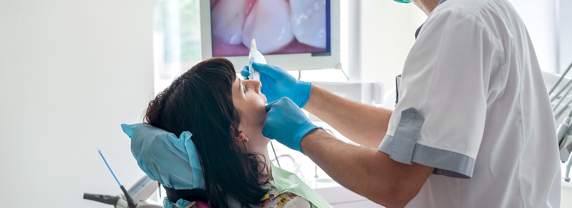

An intraoral camera is a compact, pen-sized imaging device designed to capture clear, full-color views of the teeth, gums, and other oral structures from angles that are difficult to see with the naked eye. Unlike traditional visual inspection alone, these high-resolution images bring details into sharp focus: hairline cracks, early decay, worn restorations, and subtle changes in soft tissues become visible and assessable. For patients, that means issues can be identified earlier and monitored more reliably over time.
Because the camera records still photos and video in real time, clinicians can examine both static findings and dynamic behavior — for example, how a crown margins interface with adjacent teeth or how soft tissues respond when the cheek is retracted. The technology makes it possible to document the precise condition of a tooth or restoration at a given appointment, creating an objective baseline for comparison at future visits. This capability is especially valuable for tracking the progression or resolution of minor concerns without immediate invasive treatment.
Intraoral images also highlight details that influence clinical decisions, such as the extent of plaque accumulation at the gumline, the integrity of margins around fillings, and subtle erosive wear patterns. These visual cues guide a more tailored treatment plan and help prioritize interventions that preserve tooth structure and support long‑term oral health.
During a routine appointment, the intraoral camera is used as a complementary tool alongside tactile examination and digital radiography. After a visual exam, the clinician or hygienist gently positions the camera to capture targeted views of suspicious areas identified through probing or from patient symptoms. Images are displayed instantly on the operatory monitor, allowing the dentist to inspect both the overall field and magnified details without needing to reposition the patient repeatedly.
The process is quick, noninvasive, and completely comfortable for most patients. Because the camera captures multiple perspectives, clinicians can confirm their preliminary impressions and decide whether further testing or monitoring is appropriate. It also serves as an efficient way to document treatment progress — for instance, pre- and post-restoration images can be stored to demonstrate precisely what was addressed during the appointment.
Integrating intraoral imaging into the standard exam workflow helps the dental team maintain a consistent, evidence-based record. That consistency improves the quality of follow-up care and reduces uncertainty when evaluating subtle changes between visits.
High-quality intraoral photographs enhance diagnostic accuracy in several ways. First, they expose visual signs of disease earlier than a cursory glance might, enabling dentists to catch problems at stages when conservative measures are more likely to succeed. Second, the magnified view helps in assessing margins, contacts, and occlusal wear with more precision, which supports better-fitting restorations and improved long-term outcomes.
When developing a treatment plan, clinicians rely on objective images to communicate the rationale for recommended care. Intraoral photography assists in treatment sequencing — for example, deciding whether to monitor a lesion, perform a conservative restoration, or refer to a specialist. It also supports interdisciplinary cases by creating a shared visual record that other dental professionals can consult when coordinating care.
Because the images are saved to the patient’s electronic chart, they become part of a growing clinical archive. This longitudinal documentation is an invaluable resource when evaluating the effectiveness of interventions and making evidence-based adjustments to a patient’s preventive or restorative strategy.
One of the most immediate benefits of intraoral imaging is its ability to transform the patient experience through clear, visual communication. Rather than relying solely on verbal descriptions, clinicians can show patients exactly what they see, making complex conditions understandable at a glance. This clarity reduces confusion and helps patients feel more informed about their oral health status and the reasoning behind recommended treatments.
Visuals are particularly helpful for demonstrating areas that require improved home care, such as localized plaque retention or early enamel breakdown. When patients can see the problem, they are often more engaged in the discussion about preventive measures and daily habits that influence outcomes. The camera also helps normalize routine monitoring: showing incremental improvements or areas of stability reinforces positive behavior and encourages adherence to hygiene recommendations.
Because the images are stored in the chart, the clinical team can review changes together at follow-up visits, ensuring that patients and providers share a common understanding of progress and next steps.
Intraoral photographs are more than visual aids; they are clinical documentation that integrates seamlessly into modern dental records. Captured images can be attached to the patient’s electronic health record, annotated with clinical notes, and used to support diagnostic codes and treatment rationales. This level of documentation improves continuity of care, especially in complex cases that involve multiple providers.
When a referral is necessary — for example, to an endodontist, periodontist, or prosthodontist — intraoral images provide receiving clinicians with immediate, context-rich information before the patient’s consultation. Photographs can illustrate the precise area of concern, show the condition of adjacent structures, and communicate any restorative challenges that might affect specialist planning. That early visibility helps specialists prepare targeted evaluations and streamlines collaborative care.
Additionally, because images can be shared in standard formats, they support lab communication for prosthetics and align with the digital workflows used in modern dental laboratories. The result is more predictable, efficient treatment from diagnostics through the final restoration.
Intraoral cameras are a practical, patient-friendly technology that enhances clinical visibility, improves diagnostic confidence, and strengthens communication between clinicians and patients. By capturing detailed, objective images and storing them within the dental record, this tool supports conservative decision-making, clearer treatment planning, and seamless collaboration with specialists and laboratories.
At the office of Silk Dental Delray Beach (formerly Marc Bilodeau DMD), we incorporate intraoral imaging as part of our comprehensive approach to preventive and restorative care to ensure patients receive well-documented, thoughtful treatment recommendations. If you’d like to learn more about how intraoral photography can benefit your care, please contact us for more information.
Quick Links